Sub-Total: €0.00


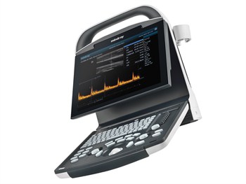

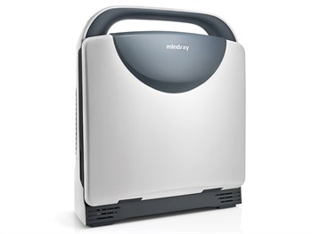
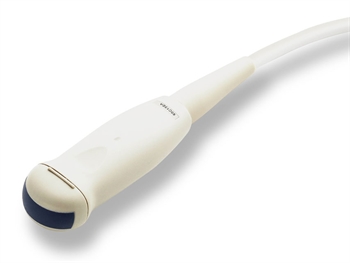
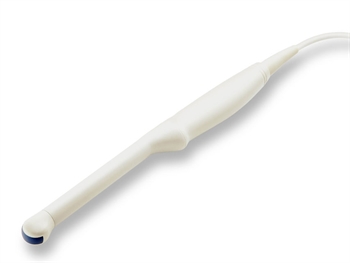
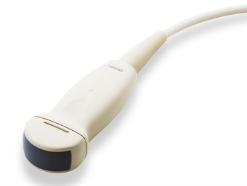
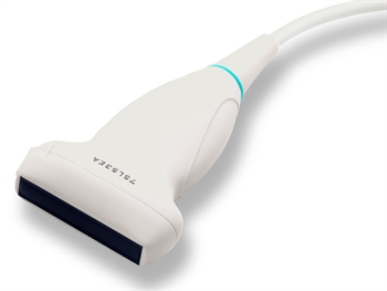
Mindray DP-10 Ultrasound System
DP-10 slim and smart, good image quality, easy to use and lightweight to take it anywhere. It combines fashionable design, diagnostic confidence and economy.
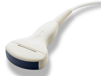
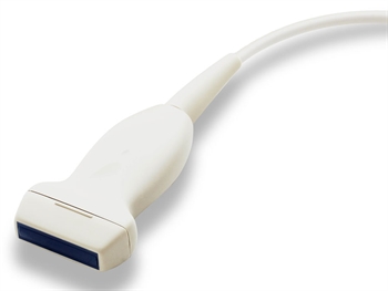
Does not include probe
You must follow the steps below:
- Select the ultrasound system and add to cart
- Select the probe(s) and add to cart
- Proceed to Cart and Checkout Page if not purchasing any additional items
€1,330.07 – €2,657.90
€1,330.07 (Excl. VAT)
€1,635.99 (Incl. VAT)
Technical Specifications:
Display modes and imaging processing:
Broadband, multi-frequency imaging: B, B/B, 4B, B/M, M
Imaging processing:
– multi-frequency probes for 2D imaging modes
– iClear: speckle reduction image
– THI (Tissue Harmonic Imaging) with convex probe 33976 only
– TSI (Tissue Specific Imaging)
– zoom function: spot zoom, pan zoom
– ExFOV (Extended field of view)
Extended view of the anatomical structure on all convex and linear probes allow to discover better diagnostic information.
– iTouch (Auto image optimization)
– colour map
User interface
– alphanumeric QWERTY keyboard with backlight
– trackball: speed presettable
– 8-segment TGC
– user defined blank keys: shortcut for easy access to menus
iStation™ intelligent workflow – Intelligent patient data management system
– integrated search engine for patient data
– detailed patient information review
– intelligent data backup/restore
– patient data/image sending, data deleting (US B , DVD, DI COM)
– exam managing: activate exam
Storage/review mode:
– image archive on hard disk and USB mobile storage medium, temporary saving in cine memory, directly transfer report to PC
– 2 USB ports
– multiple image formats: BMP, JPG, DCM, FRM, AVI, DCM, CIN
– iVision: demo player
– Cine review: auto, manual (auto review segment can be set), supports linked cine review for 2D, M images
– Cine memory capacity (max.)
– clip length presettable: 1-60 s
B mode: 11959 frame
M mode: 110.0 s
Supplied with GB, IT manual and CD manual (GB, FR, IT, ES, PT, DE, RU, BR).
iScanHelper function – Dedicated inbuilt tutorial function to enhance ultrasound experience.
– wide selection of application specific exam planes
– anatomical illustrations
– standard ultrasound images
– scanning reference pictures
– tips on scanning and diagnosing
Optional PW doppler for probes 33976 (Convex), 33977 (Linear)
DP-10 support superb PW Doppler imaging to access blood flow in different clinical conditions.
With PW, doctors will be able to:
– differentiate non echoarea and blood vessels
– distinguish arteries and veins
– evaluate blood flow velocity for more comprehensive diagnosis
– Check the health of a fetus by blood flow evaluation of the umbilical cord
Features:
Parameter adjustment: Gain, Sample volume size, Scale, Steer, Dynamic range, Wall filter, etc.
Measurement: Trace, HR, Velocity, PS/ED, PI, RI, etc.
Related Documents:
You May Also Like

Bosch

Littmann

Saver One

Omron

Omron

Iem

Braun

and

riester

mesi

medi Plent

sono site

wench allyn

plinth medical
Clinell
